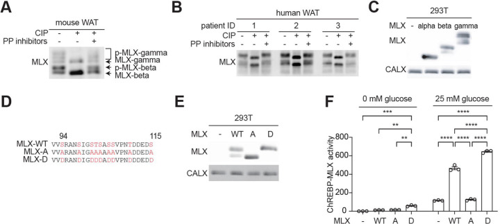
Fig. 1. MLX phosphorylation promotes ChREBP-MLX activity.
a-b. MLX phosphorylation in mouse (a) and human (b) white adipose tissue (WAT) lysates treated with calf intestine phosphatase (CIP) or CIP and phosphatase (PP) inhibitors. n=3.
c. MLX phosphorylation in 293T cells expressing MLX isoforms alpha, beta, or gamma. CALX serves as a loading control. N=3.
d. Amino acid sequence alignment of wild-type MLX (MLX-WT), phospho-deficient MLX-A or phospho-mimetic MLX-D.
e. MLX phosphorylation in 293T cells expressing MLX-WT, -A, or -D. N=3.
f. ChREBP-MLX luciferase reporter activity in 293T cells expressing ChREBP, HK2 and MLX-WT, -A, or -D. Cells were starved for glucose overnight and/or treated with 25 mM glucose for 3 hours. Two-way ANOVA **p<0.01, ***p<0.001, ****p<0.0001. N=3.

