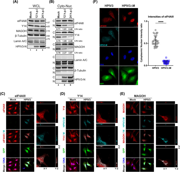
Figure 6. Subcellular localization of core EJC in HPIV3 infected HeLa cells.
(A-B) Immunoblotting analysis of whole cell lysates, cytoplasmic, and nuclear fractions from HPIV3-infected HeLa cells at 12- and 24-hours post-infection (hpi). The levels of eIF4AIII, Y14, and MAGOH were examined, with β-tubulin and Lamin A/C serving as markers for the purity of cytoplasmic and nuclear fractions, respectively. The ratios below the blots indicate the relative intensities of eIF4AIII, Y14, and MAGOH from the cytoplasm to the nucleus. (C-E) XYZ planes of 3D confocal micrographs depicted HeLa cells at 24 hours post-infection with HPIV3 at m.o.i of 5. Cells were fixed and stained with (C) anti-eIF4AIII, (D) anti-Y14, or (E) anti-MAGOH antibodies (red), and anti-HPIV3-M antibody (cyan) to label the viral matrix protein. Nuclei were counterstained with Hoechst (blue), and GFP fluorescence indicates HPIV3 infection. Enlarged orthogonal projections of the infected cells (white dashed line) are shown on the right, displaying the EJC protein, HPIV3-M, and the merged channels. Scale bars represent 20 μm. (F) Left: HeLa cells infected with either HPIV3 or HPIV3ΔM at an m.o.i. of 5 were fixed at 24 hours post-infection and stained with anti-eIF4AIII (red), anti-HPIV3-M antibodies (cyan), Hoechst for nuclei (blue), and GFP fluorescence indicates HPIV3 infection. Representative fields of cells for each condition are shown. Right panel: Quantification of cytoplasmic/nuclear eIF4Alll intensity (C: N) ratios was performed on 30 individual cells, as described in Materials and Methods. Statistical significance was analyzed by unpaired t-test. **** P <0.0001.

