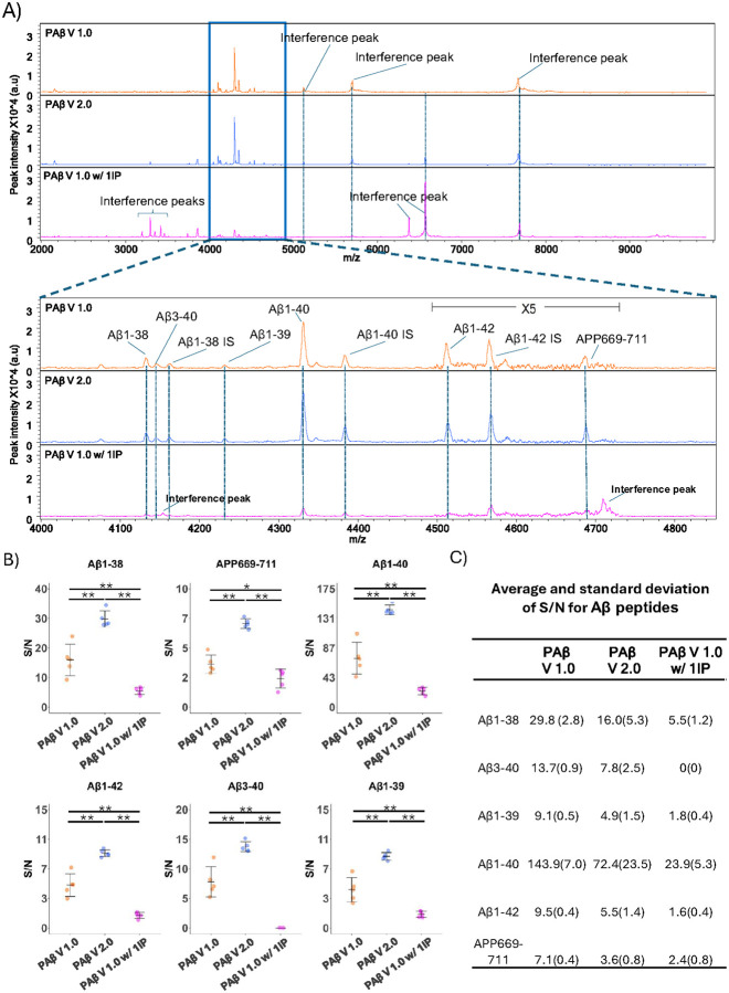
Figure 3. The spectra of IP-MS assays with S/N comparison.
(A) MALDI–TOF mass spectra of Aβ peptides derived from plasma replicates utilizing the PAβ V1.0 assay, 10% N4PE CSF diluent (PAβ V2.0 assay) and PAβ V1.0 assay with 1IP. Representative spectra from each experiment are presented. Interference peaks were consistently observed at 5771.1 m/z and 7746.8 m/z across all assays. Additionally, another interference peak at 6631.0 m/z was consistently noted in all assay formats except the PAβ V1.0 assay. Interference peaks at 3200 m/z to 3500 m/z and 6432.4 m/z were observed in PAβ Vi.0 assay with 1IP only. Upon magnification to the range of 4000–4850 m/z, the theoretical m/z values of peptides are as follows: 4i32.6 m/z for Aβ1–38, 4i44.7 m/z for Aβ3–40, 4231.8 m/z for Aβ1–39, 4330.9 m/z for Aβ1–40, 45i5.i m/z for Aβ1–42, and 4689.4 m/z for APP669–711. Aβ1–38 IS at 4160.7 m/z, Aβ1–40 IS at 4383.3 m/z, and Aβ1–42 IS at 4569.3 m/z were utilized as internal standards for the normalization of mass spectra. Notably, an interference peak was detected at 4153.4 m/z in samples processed using PAβ V1.0 assay with 1IP but not in the other assays. (B) S/N ratios were compared across three assays in triplicates, with asterisks indicating significant differences (*p < 0.05, **p < 0.0i) as determined by the Wilcoxon Rank Sum test. (C) The averages and standard deviations of the S/N ratios are listed.

