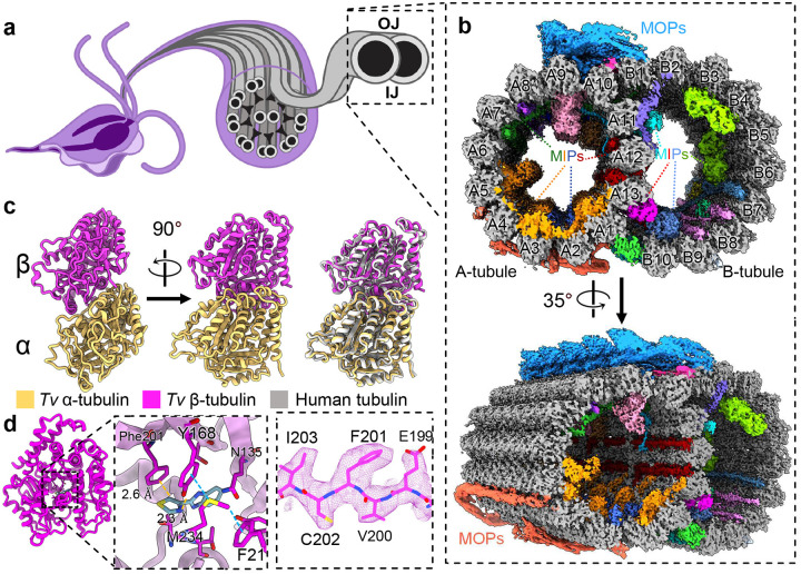
Figure 1. Cryo-EM reconstruction of the doublet microtubules from Tv.
(a) Diagram of axoneme from the flagella of T. vaginalis. (b) Cross-section of Tv-DMTs with microtubule inner proteins (MIPs) and microtubule outer proteins (MOPs) indicated with various colors. A- and B-tubules, as well as protofilaments, are labeled. (c) Atomic models of α and β tubulin, superimposed with human tubulin (right). (d) Alternate view of Tv β tubulin (left) and docked thiabendazole molecule (blue) fit into putative binding site with adjacent residues shown (right) with cryo-EM map density. IJ: inner junction; OJ: outer junction.

