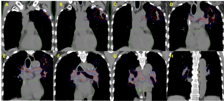
Figure 3. Planning CT contours of the left lung and mediastinal non-small cell lung cancer (NSCLC).
Coronal planning CT slices with contours display gross tumor volume (red), planning target volume (blue), heart (green), trachea and large bronchus (light blue), esophagus (orange), and bronchus/small airway (pink). Slices are shown every 1 cm and go from the anterior to the posterior from the (A) anteriormost slice, showing the anterior extent of the disease, which extends towards the second and third ribs in the left lung. Additional posterior slices (B, C) reveal bulky left lung and left mediastinal disease, and slices (D-G) demonstrate the disease extending to the right mediastinum. The most posterior CT slice (H) depicts the posterior extent of the planning target volume, with no additional gross tumor volume.

