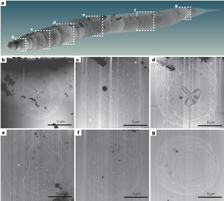
Fig. 3. Lamella TEM overviews sample the anatomy of a C. elegans L1 larva along the anterior–posterior body axis.
Native tissue scattering contrast is sufficient to extract a considerable amount of anatomical information from low-magnification TEM overviews of the lamellae generated in the double-sided-attachment Serial Lift-Out experiment. a, Schematic representation of 29 body transverse-section overviews obtained from the final lamellae along the anteroposterior body axis of an L1 larva. Anterior is located to the left; posterior is to the right. The OpenWorm project model of the adult C. elegans worm was used for illustrating the possibility of back mapping, as a cellular model of the L1 larva only exists for its head and not the entire body39. The cross-sections were cropped from lamella-overview images and mapped back to a position derived from the known sectioning distance and anatomical features discernible in the corresponding cross-section. Dashed white frames indicate overviews with corresponding magnified representations in b–g. b, This lamella originates from ~15 µm along the anterior–posterior axis. Clearly visible are the three lobes of the anterior pharyngeal lumen in the center of the worm cross-section (asterisk) and the relatively electron-dense pharyngeal lining. c, Overview from the anterior part of the pharyngeal isthmus ~42 µm along the anteroposterior axis. Note the nerve ring (dashed line) surrounding the central pharynx. Additionally, the alae (asterisks) running along the left and right lateral sides of the worm become obvious. d, Overview of a lamella of roughly the center of the posterior pharyngeal bulb region. The central grinder organ is clearly discernible (asterisk). This section is positioned ~65 µm along the anterior–posterior axis. e, A section roughly mid-body. The intestinal lumen (dashed line) and intestinal cells (dotted line) are obvious. The darker cell slightly left of the body center is likely one of the gonadal primordial cells (asterisk). The section is from ~115 µm along the anteroposterior axis. f, In this mid-body section, the intestinal lumen (dashed line) can again be clearly discerned. The section can be mapped to ~132 µm along the anteroposterior axis. g, Section showing the intestinal lumen at ~155 µm along the anteroposterior axis. The data presented in this figure stem from experiment 2.

