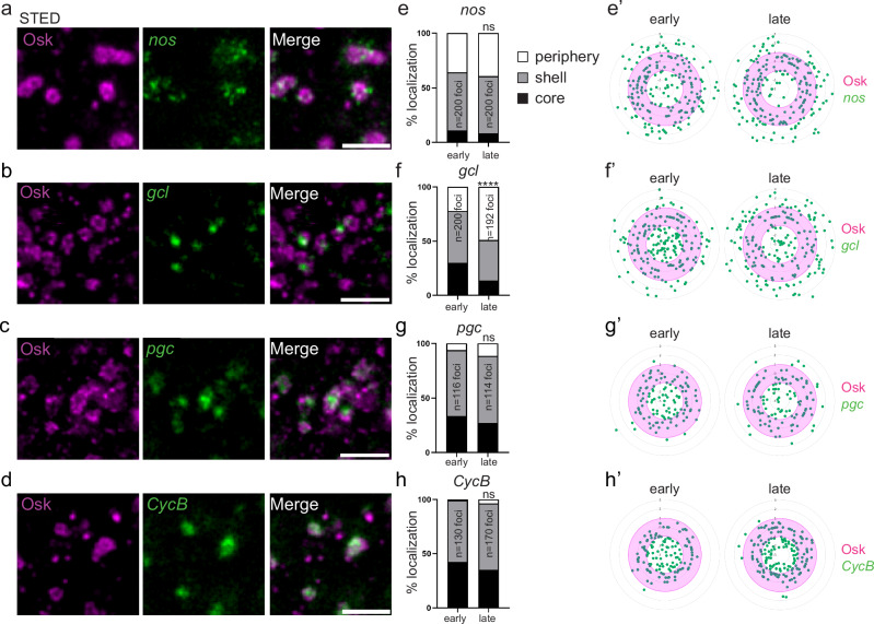
Fig. 4. mRNA localization within germ granules.
a–d STED images of immuno-smFISH of wild-type embryos (20–120 min) with anti-Osk (magenta) and smFISH probes (green) against nos (a), gcl (b), pgc (c), and CycB (d) mRNAs. e–h Percentage of localization of nos, gcl, pgc, and CycB mRNA foci in early (0–20 min) and late (20–90 min) wild-type embryos, in the core (black), in and at the surface of the shell (grey) and at the immediate periphery (white) of germ granules from images as in (a–d). ns: non-significant, ****p < 0.0001 using the χ2 test. p = 0.61 in (e), 1.49 × 10⁻¹⁸ in (f), 0.26 in (g) and 0.17 in (h). e’–h’, Radar plots of the relative localization of nos, gcl, pgc, and CycB mRNAs (green dots) within Osk immunostaining (magenta) in early (0–20 min) and late (20–90 min) embryos from images as in (a–d). The granule shell is in pink. Scale bars: 1 µm. Source data are provided as a Source Data file.

