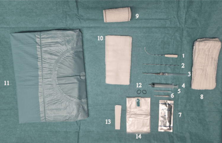
Figure 1. Material needed to perform the surgery.
The probe cover (14) was attached to the probe with elastic rubber bands (12) and fixed on the sterile drape with tapes (13).
A sterile ultrasound gel (7) is applied between the probe cover and the patient skin.
The patient is positioned in the supine position with the operated hand placed on a table and stabilized with two abdominal sponges (8) in the neutral position of the wrist. An 18G needle (6) is used to harvest the lidocaine in a 10cc syringe (4) and a 20 G needle (5) for injection through the entry point.
A 14G catheter (3) is then introduced through the skin and the forearm fascia to increase the entry point size and replaced by the round-ended stylus (2) to palpate the TCL and define the cut trajectory. After the stylus removal, the cutting instrument (1) is introduced through the entry point to perform the antegrade release.
After the release, the round-ended stylus is used to palpate the ligament and be sure of the completeness of its release. If this is not the case, the exact location of the remaining fibers is defined by probe palpation, and the cutting instrument is re-introduced to complete the release.
Three 10x20 sponges (10) are then used to make a compressive dressing with a Velpeau band (9) that will be removed by the patient after 12 hours.

