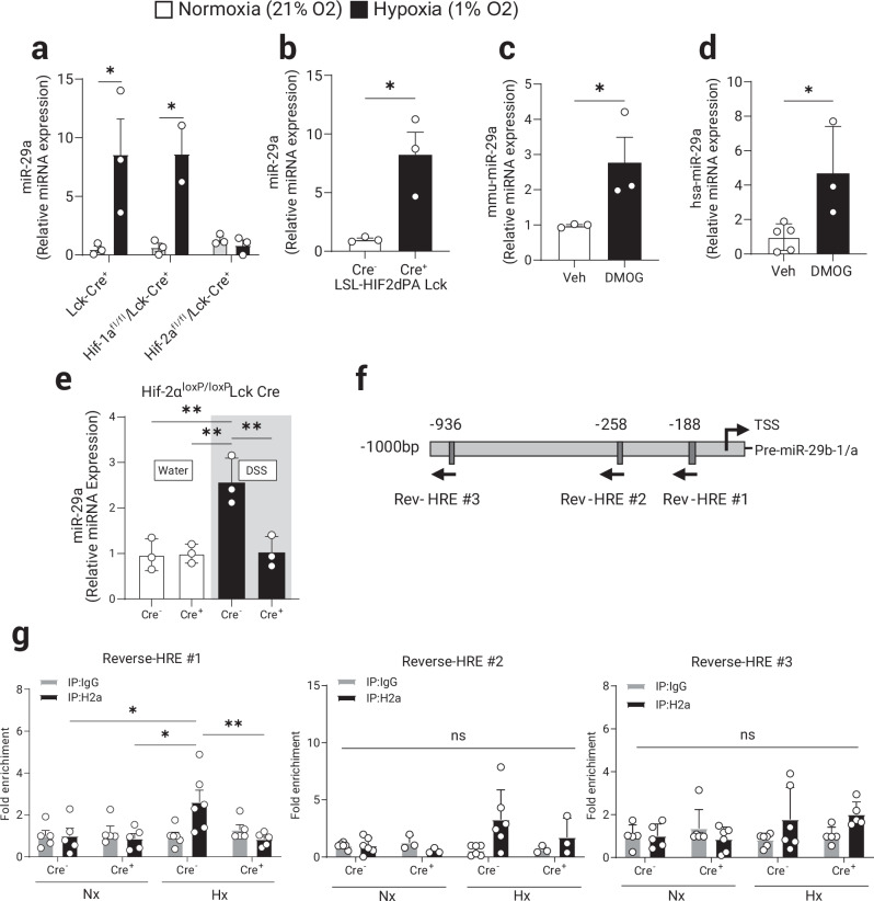
Fig. 3. HIF-2α mediates the induction of miR-29a in CD4+ T cells during hypoxia.
Primary mouse naïve CD4+ T cells were isolated from spleens and MLN of control (Lck-Cre+) or mice with a selective HIF-1α or HIF-2α deficiency in T cells. Isolated CD4+ T cells were cultured ex vivo for 8 h stimulation with TCR (anti-CD3/28) in either normoxia (21% O2) or hypoxia (1% O2). a miR-29a induction by hypoxia is absent in Hif-2α-deficient CD4+ T cells, (n = 2–3, two-way ANOVA). b miR-29a is significantly induced in primary CD4+ T cells isolated from Hif-2α-over-expressing mice (LSL-HIF2dPA Lck Cre+) cultured ex vivo for 8 h stimulation with TCR stimulation (n = 3, two-tailed T test). Pharmacologic stabilization of HIF-2α with DMOG treatment (1 mM) induced miR-29a in: (c) primary CD4+ T cells from WT (B6) mice (n = 3, one-tailed T test) and, (d) CD4+ T cells isolated from PBMCs of healthy human volunteers (n = 3–5, two-tailed T test). Hif-2αloxP/loxP Lck Cre+ and Hif-2αloxP/loxP Lck Cre− mice were treated with two rounds of 3% DSS in drinking water for 5 days with 2 weeks of water in between treatments to induce chronic colitis. e Quantification of miR-29a expression in the total mouse colon tissue, (n = 3, two-way ANOVA). f Schematic representation of murine miR-29b1-a promoter with 3 most proximal putative Hypoxia Responsive Elements (HRE) indicated. More distant putative HREs (4) were omitted. g Chromatin Immuno-Precipitation (ChIP) was performed to assess in vivo DNA-protein interactions at the proximal promoter sequences in primary CD4+ T cells from Hif-2αloxP/loxP Lck Cre+ and Cre- mice cultured ex vivo for 8 h with TCR stimulation (anti-CD3/28) in either normoxia (21% O2) or hypoxia (1% O2) as per Methods (n = 6, two-way ANOVA). Male and female mice used in (a–d, g), male mice used in (e). Data expressed as Mean ±S.E.M from 2 polled independent experiments., *p < 0.05, **p < 0.01.

