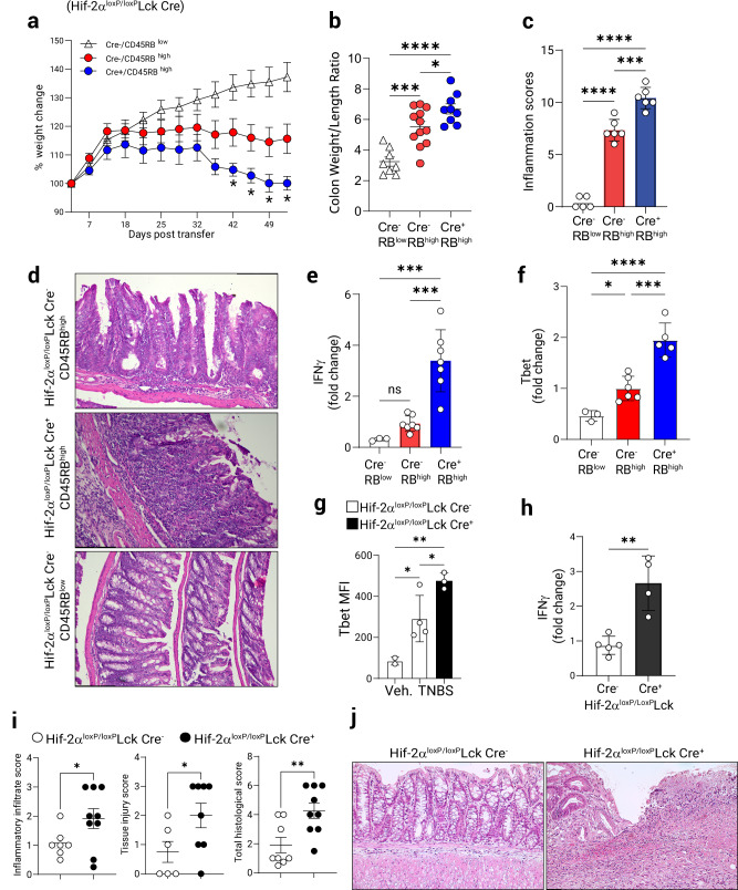
Fig. 5. T cell-intrinsic HIF-2α regulates T cell function in Colitis.
Chronic T cell-mediated colitis was induced in B6.B6.RAG1−/− mice by intraperitoneal adoptive transfer of naïve CD4+ CD45RBhigh or CD45RBlow T cells (0.5 × 10⁶/ animal) derived from Hif-2αloxP/loxP Lck Cre+ or Cre− animals. a Weight change during the course of colitis (n = 8–10, 2-way ANOVA, Cre- vs. Cre+). b Colon weight/length ratio at fulminant disease (n = 9–12, one-way ANOVA). c Histological preparations of colons stained with H&E were scored for total inflammation (as per Methods, (n = 5–6, one-way ANOVA). d Representative micrographs of colons at day 56 post-transfer, scale bar: 131.4 mm. Mesenteric lymph node CD4+ T cells were purified from adoptively transferred mice and total RNA was assayed by Q-PCR for: e IFNγ mRNA expression (n = 3–7, one-way ANOVA). f T-bet mRNA expression. Gene expression in (e, f) was normalized to 18s rRNA. (n = 3–6, one-way ANOVA). TH1 colitis was induced in Hif-2αloxP/loxP Lck Cre+ or Lck Cre+ control mice by the epicutaneous skin sensitization and subsequent rectal gavage with TNBS. At the time of necropsy mesenteric node lymphocytes were assessed by flow cytometry. g T-bet mean fluorescence intensity (MFI) in CD4+ T cells (gated on live CD3+, n = 3–4, one-way ANOVA). h IFNγ expression in mesenteric node lymphocytes was assayed by Q-PCR (n = 4–5, two-tailed T test). i Histological indices including leukocyte infiltration, tissue injury and total inflammation in the course of colitis (n = 6–9, one-tailed T test). j Representative micrographs from Lck Cre+ controls or Hif-2αloxP/loxP Lck Cre+ mice 7 days post-challenge, scale bar 100 mm. Male recipients and mixed male/female donors used in (a–f), male and female mice used in (g–j). Data expressed as Mean ± S.E.M, *p < 0.05, **p < 0.01, ***p < 0, .001. vs. indicated.

