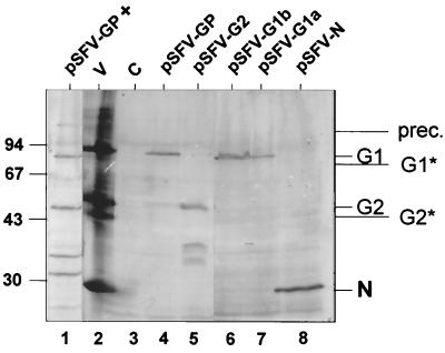
FIG. 2.
Western blots using antiserum against TSWV particles, showing the expression (21 h.p.t.) of the TSWV glycoproteins (pSFV-GP), G2 alone (pSFV-G2), G1 alone (pSFV-G1a and pSFV-G1b), and N (pSFV-N) in BHK cells. Expression of the TSWV glycoproteins in the presence of tunicamycin is shown in lane pSFV-GP + G1* and G2* indicate unglycosylated forms of G1 and G2, and prec. is the glycoprotein precursor protein. The positions of size markers are indicated on the left (in kilodaltons). V, purified TSWV particles; C, control (mock-transfected) cells.

