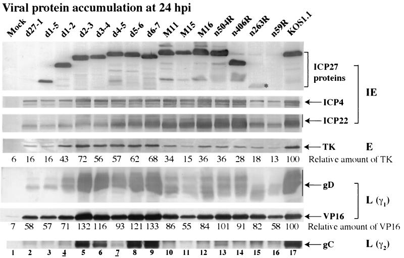
FIG. 6.
Immunoblot detection of viral protein accumulation in infected HEp-2 cells. Whole-cell extracts prepared at 24 h p.i. were used for immunoblot analyses with anti-ICP27, anti-ICP4, and anti-ICP22 (IE proteins), anti-TK (E protein), anti-VP16 and anti-gD (L proteins, γ1), and anti-gC (L protein, γ2) antibodies. Relative amounts of TK and VP16 were normalized to the amount of α tubulin and calculated as described in Materials and Methods. An asterisk marks n263R ICP27 protein since its accumulation is at low levels. Lanes 4 and 7 are underlined to mark viruses analyzed in Fig. 7.

