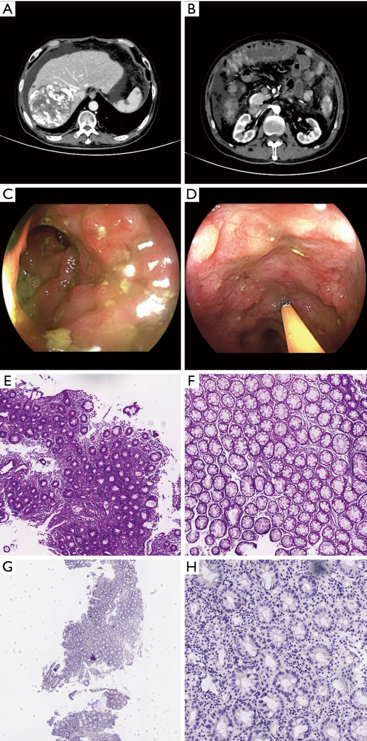
Figure 2.

Imaging and pathological features of immune checkpoint inhibitor-induced enterocolitis. (A,B) Contrast-enhanced abdominal computed tomography showing complete necrosis of the hepatocellular carcinoma, moderate perihepatic effusion and mucosal edema of the transverse colon. (C,D) Colonoscopy revealed that the total colon mucosa, especially the descending colon and transverse colon, was congested, edematous, erosive, and covered by fused shallow ulcers, which had partially swollen surface pus. (E,F) Histology confirmed active colitis with a mixed inflammatory infiltrate in the descending colon and sigmoid colon (hematoxylin-eosin staining, ×100, ×200). (G,H) Immunohistological examinations showed cytomegalovirus 2 immunonegative (immunohistochemistry, ×40, ×400).
