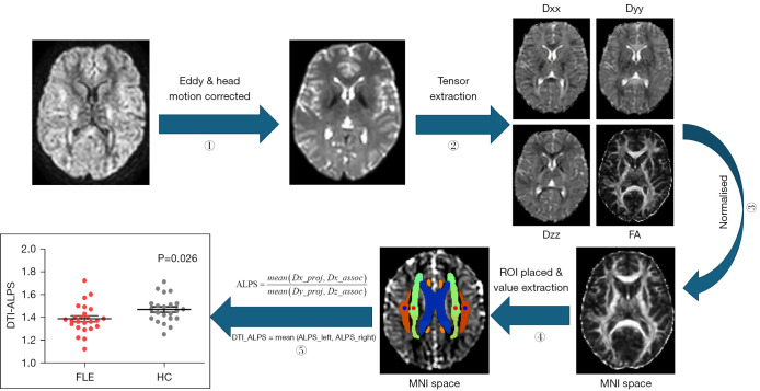
Figure 2.
Data processing and DTI-ALPS index calculation process. ① All DTI images were eddy and head motion corrected in FSL toolbox; ② FA maps and diffusivity maps of each subject were then obtained using FSL software in the direction of the x-axis (right-left; Dxx), y-axis (anterior–posterior, Dyy), z-axis (inferior superior, Dzz). ③ The ICBM DTI-81 Atlas was used as a reference to normalize the FA map and triaxial diffusivity to MNI space. ④ Four spherical ROIs were placed on the projection fibers and association fibers in the uppermost layer of the lateral ventricle body. ⑤ The mean of the bilateral DTI-ALPS-index represented the overall glymphatic function of the subjects. FA, fractional anisotropy; MNI, Montreal Neurological Institute; ROI, region of interest; ALPS, analysis along the perivascular space; DTI, diffusion tensor imaging; FLE, frontal lobe epilepsy; HC, healthy control; FSL, FMRIB Software Library.

