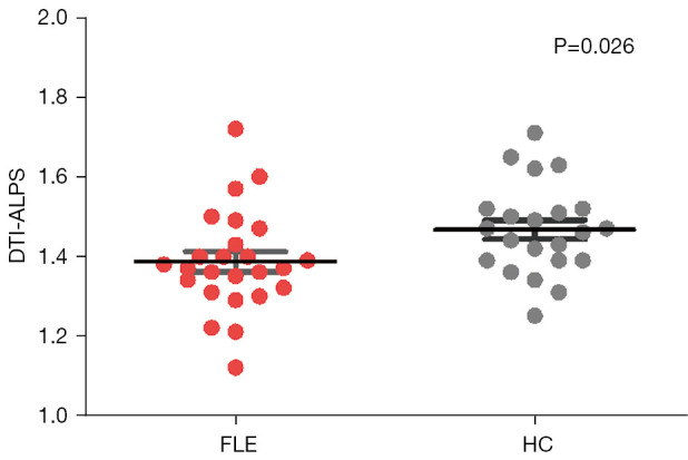
Figure 3.

Comparison of DTI-ALPS index between FLE and HC groups. The error bars indicated standard deviation. DTI-ALPS, diffusion tensor imaging analysis along the perivascular space; FLE, frontal lobe epilepsy; HC, healthy control.

Comparison of DTI-ALPS index between FLE and HC groups. The error bars indicated standard deviation. DTI-ALPS, diffusion tensor imaging analysis along the perivascular space; FLE, frontal lobe epilepsy; HC, healthy control.