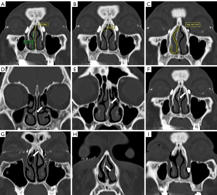
Figure 2.
Anatomic characteristics measured and evaluated by computed tomography. (A) The angle of NSD was measured on coronal CT images as the angle between the most deviated point of the septum and the midline. The height of NSD was the vertical distance from the apex of NSD to the floor of the nasal cavity and was measured parallel to the midline (green line). (B) The MNPW on the surgical side was measured in the coronal plane by selecting the level with the largest lacrimal sac visualization and was defined as the distance between the frontal processes of maxilla and the nasal septum on the operated side. (C) The minimal cross-sectional intranasal areas of the surgical passage were identified at the surgical region and then outlined and calculated. (D-H) The modified Guyuron classification of septal deformities: (D) septal tilt and localized spicule, (E) S-shaped anteroposterior and cephalocaudal septal deviations, (F) C-shaped anteroposterior and cephalocaudal septal deviations, (G) concha bullosa on the surgical side, and (H) inferior turbinate hypertrophy on the nonsurgical side (white arrows). (I,F) Images from the same patient with bilateral PANDO; the right eye was the first to be treated with En-DCR in combination with septoplasty. (I) Image obtained during the preoperative examination of this patient’s left eye, which indicated a patent anastomosis and a well-corrected septum in the right eye after surgery. NSD, basal septal deviation; CT, computed tomography; MNPW, middle nasal passage width; PANDO, primary acquired nasolacrimal duct obstruction; En-DCR, endonasal endoscopic dacryocystorhinostomy.

