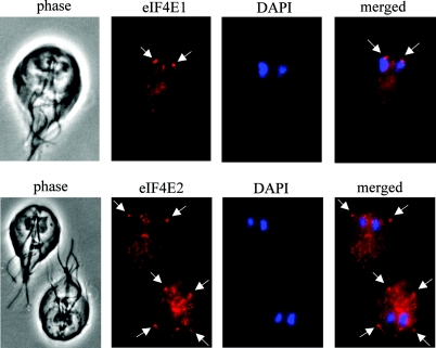
FIG. 7.
Immunofluorescence localization of eIF4Es in Giardia. Giardia WB cells stained with DAPI were further stained with rabbit polyclonal antibodies against Giardia eIF4E1 (top panel) and eIF4E2 (bottom panel). The stained cells were examined with a fluorescence microscope. Arrows point to concentrated localizations of the protein.

