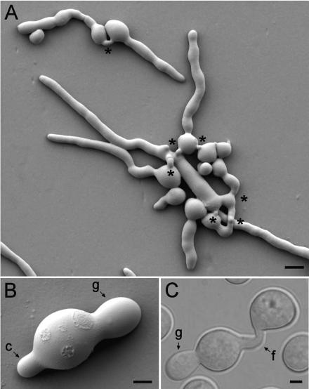
FIG. 1.
CATs and germ tubes. (A) Germinated macroconidia with long germ tubes avoiding each other and CATs which have grown (homed) toward each other and fused (asterisks). The image was obtained by low-temperature scanning electron microscopy. Bar, 10 μm. (B) A single macroconidium which has produced a germ tube (g) and a CAT (c). The image was obtained by low-temperature scanning electron microscopy. Bar, 2.5 μm. (C) Fusion of CATs (f) produced directly from two conidia, one of which has produced a germ tube (g). This is a differential interference contrast image. Bar, 2.5 μm.

