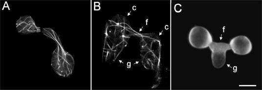
FIG. 7.
(A) Microtubules (labeled with β-tubulin-GFP) longitudinally organized and extending towards CAT tips of macroconidia which are homing toward each other. (B) After fusion, microtubules (labeled with β-tubulin-GFP) have extended through the fused CATs (f) from one macroconidium (c) bearing a germ tube (g) into the other and vice versa and, as a result, have become intermixed. (C) Germ tube (g) which is growing out of fused CATs. Images were obtained by confocal microscopy. Bar, 5 μm.

