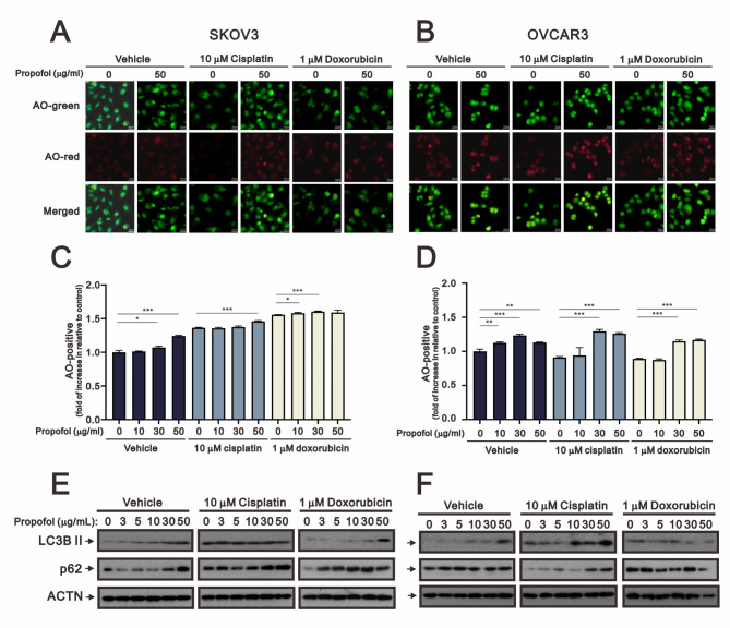
Fig. 5.
The effects of propofol in combination with cisplatin and doxorubicin on autophagy in human ovarian cells. (A-B) Fluorescence microscopy images of SKOV3 and OVCAR3 cells treated with propofol at concentrations of 0 and 50 µg/ml, and stained with AO, are shown (scale bar: 10 μm). (C-D) SKOV3 and OVCAR3 cells treated with propofol at concentrations of 0, 10, 30, and 50 µg/ml combined with either 10 µM cisplatin or 1 µM doxorubicin for 24 h were analyzed using flow cytometry with AO staining. The bars represent the mean ± SD of three independent experiments. Statistical significance is indicated by * for p < 0.05, ** for p < 0.01, and *** for p < 0.001, determined using Student’s t-tests. Cell lysates of (E) SKOV3 and (F) OVCAR3 were subjected to Western blot analysis using antibodies against the indicated proteins. ACTN was the protein loading control

