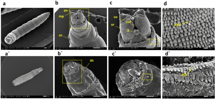
Figure 5.
M. domestica L3 scanning electron photomicrographs: (a) Displays the control larva, while (a') illustrates a shrunken larva from the CdS NPs-treated group. Moving to (b), it shows the anterior end of the 3rd larval instar with standard body details, including anterior spiracles (as), maxillary palpus (mp), and antennal complex (an). In contrast, (b') depicts the anterior end of a larva treated with CdS NPs, indicating the degeneration of the antennal complex, maxillary palpus, and anterior spiracle. (c) Presents a higher magnification of the inset in (b), showing the control facial mask with details of cutaneous teeth (cut), oral ridges (or), labial lobe (ll), and ventral organ (vo). Meanwhile, (c') shows a higher magnification of the inset in (b'), displaying the anterior end of the treated larva with extreme deformation of the antenna, maxillary palpus, and labial lobe. Finally, (d) illustrates a higher magnification of the inset in (c) of the control larva with an organized anterior spinose band (asb), and (d') depicts a higher magnification of the inset in (c') of the treated group with an unorganized, damaged anterior spinose band. Abbreviations: as = anterior spiracles; mp = maxillary palpus; an = antennal complex; cut = cutaneous teeth; or = oral ridges; ll = labial lobe; vo = ventral organ; s = spinules.

