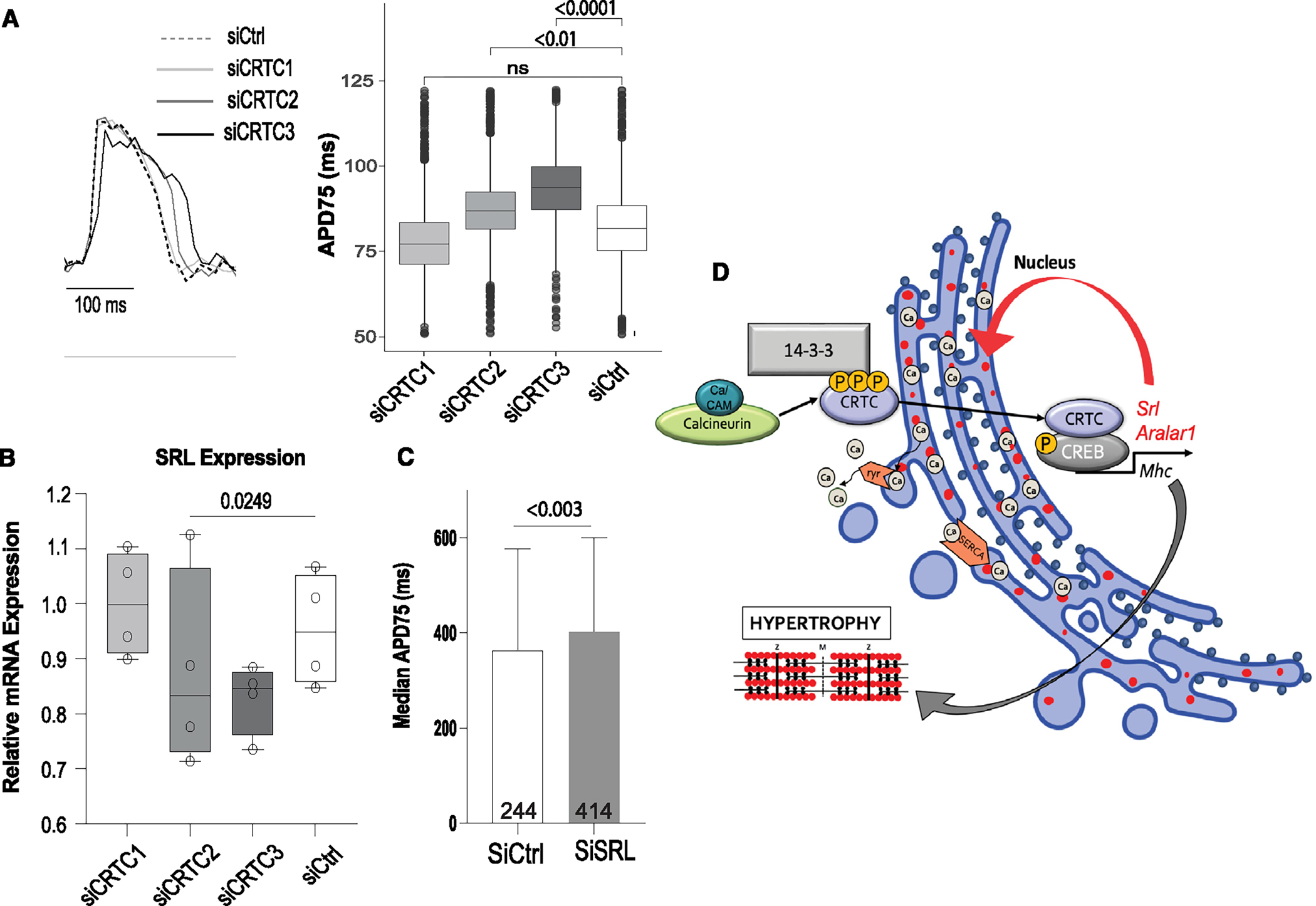
Figure 7. CRTC affects cardiac function in hiPSC-cardiomyocytes.

(A) Representative voltage traces from control siRNA- and siCRTC-transfected cardiomyocytes. APs were recorded optically from individual cardiomyocytes. APD at 75% of repolarization (APD75) was significantly increased with siRNA-mediated KD of CRTC2 and CRTC3. (Plots show max, min, median, and p values; siCtrl n = 1,338, siCRTC1 n = 1,459, siCRTC2 n = 1,427, siCRTC3 n = 1,326; significance by one-way ANOVA with Tukey’s multiple comparisons post hoc test).
(B) RT-qPCR quantification showing reduction in SRL expression in iPSC-CMs after siRNA CRTC KD. (Significance by one-way ANOVA with Tukey’s multiple comparisons post hoc test).
(C) siRNA KD of SRL prolongs APD75 in hiPSC-CMs (unpaired t test).
(D) Model of proposed CaN-CRTC-SRL signaling pathway.
