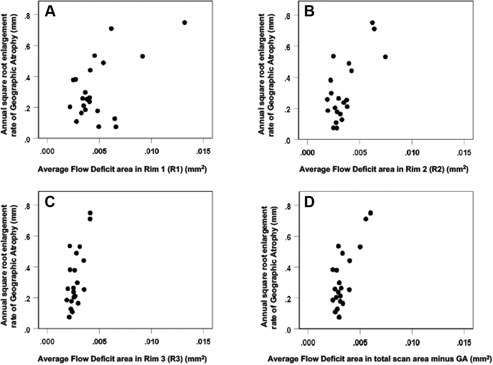
Figure 6.

Scatter plots comparing the choriocapillaris (CC) average flow deficit area (FDa) measurements (mm2) and the annual square root enlargement rates (ERs) of geographic atrophy (GA) for the 22 eyes. (A) Rim 1, (B) Rim 2, (C) Rim 3, and (D) Total scan area minus GA. The correlation between the FDa (mm2) and the annual square root ERs of GA was higher in (C) Rim 3 (Pearson r = 0.69 P < 0.001) and in (D) the area minus the GA only (r = 0.75 P < 0.001) than in (A) Rim 1 (r = 0.54 P = 0.009).
