Figure 4.
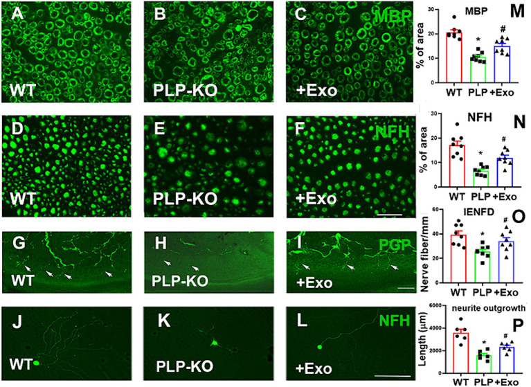
SC-Exo rescue the SC Dicer-ablation-induced morphometric changes of sciatic nerves in PLP-cKO mice. (A–F) Show representative images of MBP (A-C, green) and NF-H (D–F, green) immunoreactive sciatic nerves and PGP immunoreactive intraepidermal nerve fibers (IENF) in the hind plantar paw skin (G–I) from WT mice treated with saline (A,D,G, WT), PLP-cKO mouse treated with saline (B,E,H, PLP-KO), and PLP-cKO mouse treated with SC-Exo (C,F,I, +Exo) 8 weeks after treatment. The histogram represents the quantitative data of the percentage of MBP (M) and NF-H (N) immunoreactive area and IENF density (O). n = 8 mice/group. Scale bar in F = 25 μm, I = 50 μm. (J–L) Show that representative microscopic images of neurofilament H (NFH) + neurite of DRG neurons derived from WT mice (WT, J), PLP-cKO mice (PLP-KO, K), PLP-cKO mice treated with SC-Exo (+Exo, L). (P) Shows quantitative data of neurite length of DRG neuron under different culture conditions. Scale bar in L = 50 μm. n = 6 mice/group. One-way ANOVA followed by Tukey’s multiple comparisons tests were used. *p < 0.05 versus WT treated with saline. #p < 0.05 versus PLP-cKO treated with saline. Error bars indicate the standard error of the mean.

