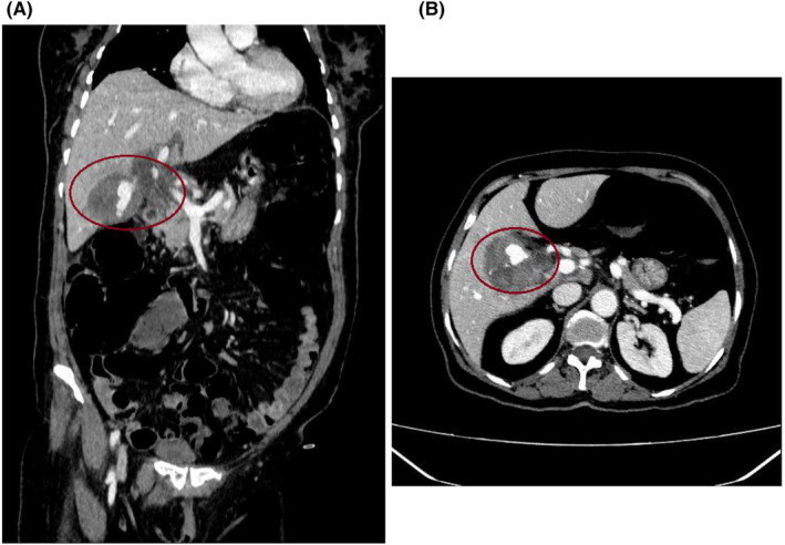
FIGURE 1.

Contrast‐enhanced computed tomography scan of the abdomen. (A) In the coronal plane, an intra‐cholecystic pseudoaneurysm of the cystic artery with a hematoma around it was shown (red circle). (B) In the axial plane, a cystic artery pseudoaneurysm with surrounding hematoma was illustrated (red circle).
