FIGURE 2.

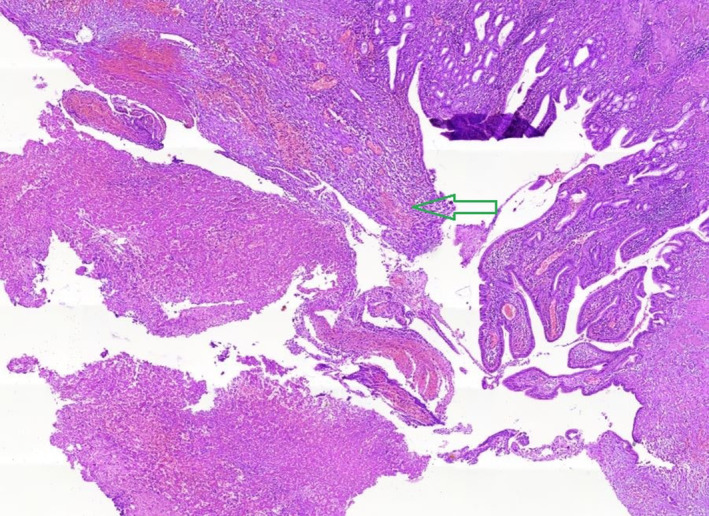
Hematoxylin and eosin (H&E) staining of the gallbladder sample demonstrated moderate infiltration along with ulcerated mucosa and the presence of polymorphonuclear neutrophils (PMNs) (green arrow) with 10x magnification.

Hematoxylin and eosin (H&E) staining of the gallbladder sample demonstrated moderate infiltration along with ulcerated mucosa and the presence of polymorphonuclear neutrophils (PMNs) (green arrow) with 10x magnification.