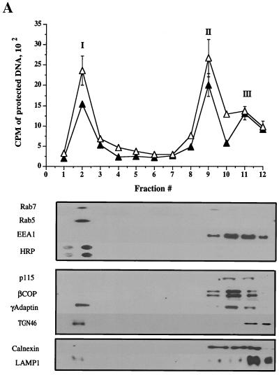
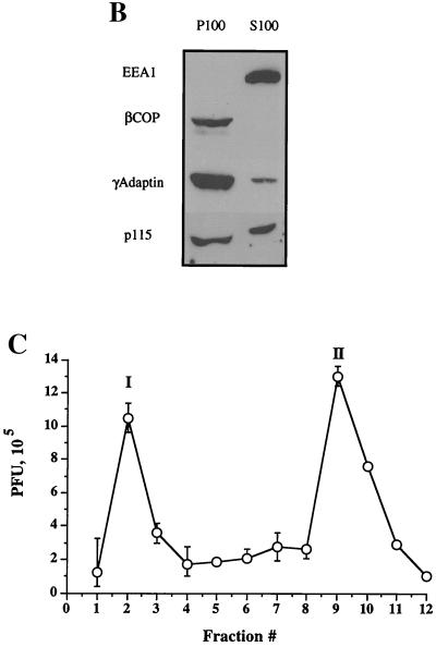
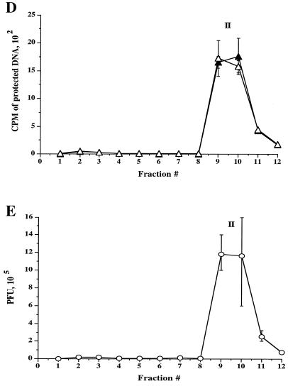
FIG. 3.
Fractionation of intracellular virus by sucrose equilibrium density gradient centrifugation. Intracellular (A and C) or extracellular (D and E) virus was prepared as described for Fig. 2 and subjected to sucrose density gradient centrifugation. Fractions were collected from the top of the gradient, as indicated on the horizontal axes (fraction 1 is at the top of the gradient). (A) Top: Intracellular fractions were assayed for packaged and enveloped DNA (open and solid symbols, respectively). The three peaks of radioactivity are labeled I, II, and III. Bottom: Intracellular fractions were subjected to Western blot analysis for the organelle markers rab7 (late endosomes), rab5 and EEA1 (early endosomes), HRP (bulk-phase endocytic compartments), p115 and β-COP (Golgi apparatus), γ-adaptin (TGN, clathrin-coated vesicles), TGN46 (trans-Golgi network), calnexin (microsomes—ER), and LAMP1 (lysosomes). (B) PNSs were subjected to a 100,000 × g spin, and the resulting pellet (P100) and supernatant (S100) were Western blotted for the antigens indicated at the left. (C) Intracellular fractions were titrated for PFU. (D) Extracellular virus gradient fractions were assayed for packaged and enveloped DNA (open and solid symbols, respectively). (E) Extracellular virus gradient fractions were titrated for PFU. All plotted values represent the mean of duplicate samples, and error bars indicate the range from the mean.


