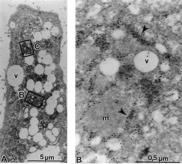
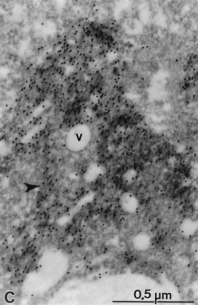
FIG. 9.
Subcellular localization of HCV proteins. Cells of cell line 5-15 were processed 4 days postseeding for immunoelectron microscopy without osmium fixation as described in Materials and Methods. The sections were probed with a mixture of polycional antisera directed against NS3, NS4B, NS5A, and NS5B, followed by 12-nm-diameter colloidal gold particles conjugated to anti-rabbit antibodies. (A) The cell in low magnification overview displays a strong vesiculation and a couple of gold-labeled areas (boxed). (B and C) Enlargements of the areas indicated by rectangles in panel A. (B) Accumulations of gold label in elongated structures representing cisternae of the ER (arrowheads). Note the scarcity of gold particles on vesicles (v), on mitochondria (m), or outside the labeled cisternae. (C) Gold-labeled cisternae are easily identified (arrowhead). Additional antibody binding could be seen on numerous submembranous structures around small vesicles (v).


