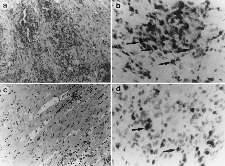
FIG. 5.
Cardiac histopathology. Tissues were sampled on day 14. In untreated control mice (left), severe myocardial necrosis (a) with MIP-2-positive cells (b) was observed. However, in anti-MIP-2 MAb (100 μg/day)-treated mice (right), myocardial necrosis (c), and MIP-2-positive cell infiltrations (d) were less severe. Arrows and arrowheads indicate cells positively stained for MIP-2. (a and c) hematoxylin and eosin staining; magnification, ×80. (b and d) MIP-2 staining; magnification, ×150.

