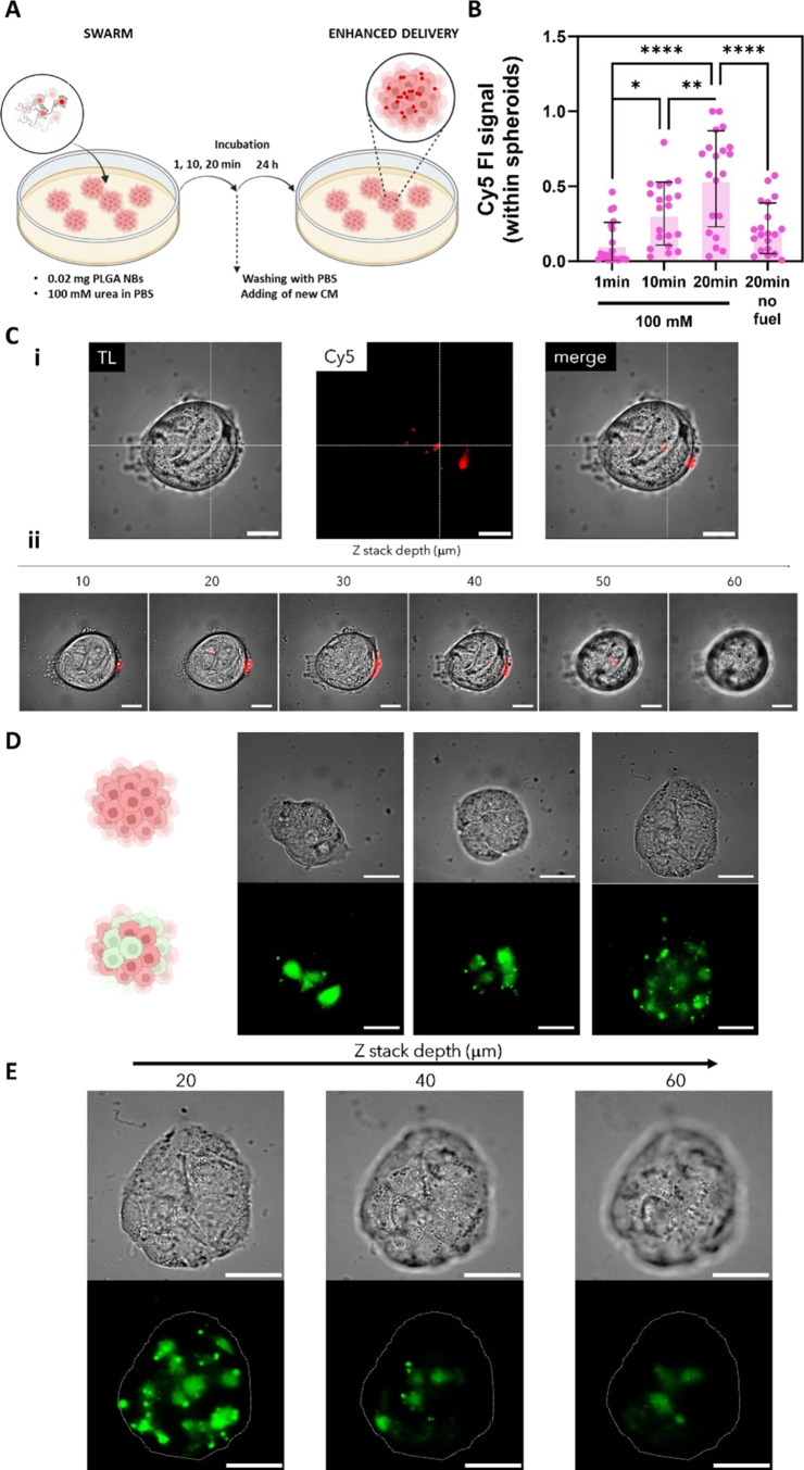
Figure 6.
Delivery and transfection by swarms of LBL PLGA NBs in spheroids of human urinary bladder-derived RT4 cells (3D). (A) Schematic overview of the experimental design for the evaluation of the delivery efficiency of swarms of LBL PLGA NBs. For swarming effect evaluation: a drop of 2 μL containing 0.02 mg of Cy5-labeled LBL PLGA NBs was added in the center of a dish previously seeded with spheroids of RT4 cells and filled with 100 mM urea in PBS. After adding the drop the system was left to evolve undisturbed for 1, 10, or 20 min. After this period, the cells were washed with PBS and were incubated for 24 h in fresh medium prior to quantifying the delivery efficiency by fluorescence microscopy. (B) Normalized mean fluorescence intensity of the NB’s signal within RT4 spheroids at different incubation times. The condition of 20 min incubation in the absence of urea was included as a control. Results correspond to at least 20 spheroids per condition. Statistical significance (one-way ANOVA, with multiple comparisons) is indicated when appropriate (*p < 0.05, **p < 0.01, ***p < 0.001, ****p < 0.0001). (C) Microscopy images of: (i) the middle focal plane (transmission light, red channel showing Cy5-labeled NBs and merged image) and (ii) z-stack (merged image) of an RT4 spheroid after 20 min treatment with 0.02 mg of Cy5 labeled NBs in 100 mM urea. The scale bar corresponds in every case to 100 μm. (D) Representative fluorescent microscopy images and (E) z-stack of a transfected RT4 spheroid expressing eGFP (green signal) 24 h after treatment with 0.02 mg of pDNA-loaded LBL PLGA NBs. The scale bar corresponds to 100 μm. Created with BioRender.com.

