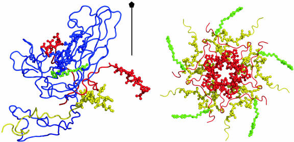
Fig. 3.
Capsid structure at the 5-fold axis. (Left) Ribbon drawing of VP1 (blue) and part of VP3 (red) and VP4 (yellow), with residues with large  shown as spheres. These residues are VP1 residues 99–109, 221, and 226; VP3 N-terminal residues 1–10 and C-terminal residues 233–236; and VP4 N-terminal residues 29–42. WIN 52084 is colored green. (Right) N-terminal residues of VP3 and VP4 from five protomers around the 5-fold rotation axis. Colors are as described for Left.

