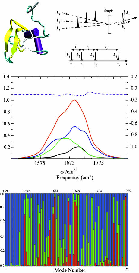
Fig. 1.
The molecular structure of TB6 and pulse sequence configuration. (Top Left) The molecular structure of TB6. The Purple sections are the helices, the yellow sections are the β-strands, and the green and white sections are randomly coiling structures. (Top Right Upper) Pulse configuration for heterodyne four-wave mixing techniques. k1, k2, and k3 are the input pulses, and ks is the signal generated, which is in the same direction as the detection pulse k4.(Top Right Lower) The pulse sequence for coherent 2D experiments. ti are the time intervals between the pulses, and τi is the peak time of pulse i. (Middle) Red, linear absorption; black, AS(α); green, AS(β); blue, AS(γ); purple dash, the difference of the additive and the actual spectrum. (Bottom) Motif content of the vibrational eigenstates of TB6. Red, α; green, β; blue, random coil. The plot is vs. mode number; the (nonuniform) frequency scale is given at the top.

