
Fig. 4.
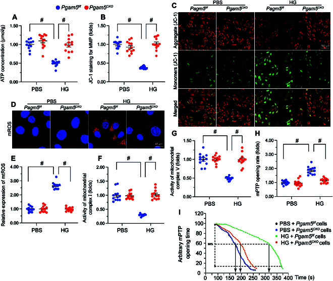
PGAM5 ablation attenuates hyperglycemia-mediated mitochondrial damage. Primary neonatal cardiomyocytes isolated from Pgam5CKO and Pgam5f/f mice were cultured under high glucose (30 mmol/l) medium for 48 h to induce hyperglycemic stress in vitro. Cardiomyocytes incubated in normal glucose (5.5 mmol/l) medium were used as control. (A) ELISA-based analysis of ATP production in primary cardiomyocytes. (B) Detection of mitochondrial membrane potential (ΔΨm) in cardiomyocytes loaded with the JC-1 probe. (C) The red-to-green fluorescence ratio was used for semiquantitative analysis of changes in ΔΨm. (D) Analysis of mtROS generation in cardiomyocytes loaded with MitoSOX Red. (E) The levels of mtROS were normalized against those of the control group. (F and G) ELISA-based determination of the activity of mitochondrial respiratory complex I (F) and complex V (G). (H) Detection of mPTP opening in cardiomyocytes loaded with TMRE. (I) Quantification of mPTP in cardiomyocytes. Values are presented as mean ± SEM from 4 independent experiments. #P < 0.05.