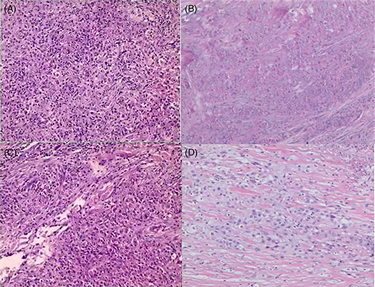
Figure 1.

Pathology of the trichilemmal carcinoma patient. (A & B) Initial postoperative pathology showed that malignant transformation of outer root sheath tumor and nests of atypical cells with keratinization and necrosis, diagnosed as trichilemmal carcinoma (TC) with parotid gland and lymph node metastasis. (C & D) The pathology of re-excision showed that there were large necrotic foci in the hyperplastic fibrous tissue, small nested atypical cells, a large amount of foam cell infiltration, confirmed metastatic TC with extensive necrosis and perineural invasion. (A & C hematoxylin and eosin ×100, B & D hematoxylin and eosin ×40).
