Figure 5.

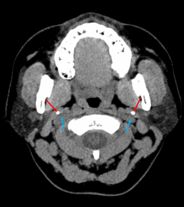
Axial view of the non-contrasted CT of the neck showing bilateral elongated styloid processes (red arrows) that were in close proximity with the internal jugular vein (blue arrows).

Axial view of the non-contrasted CT of the neck showing bilateral elongated styloid processes (red arrows) that were in close proximity with the internal jugular vein (blue arrows).