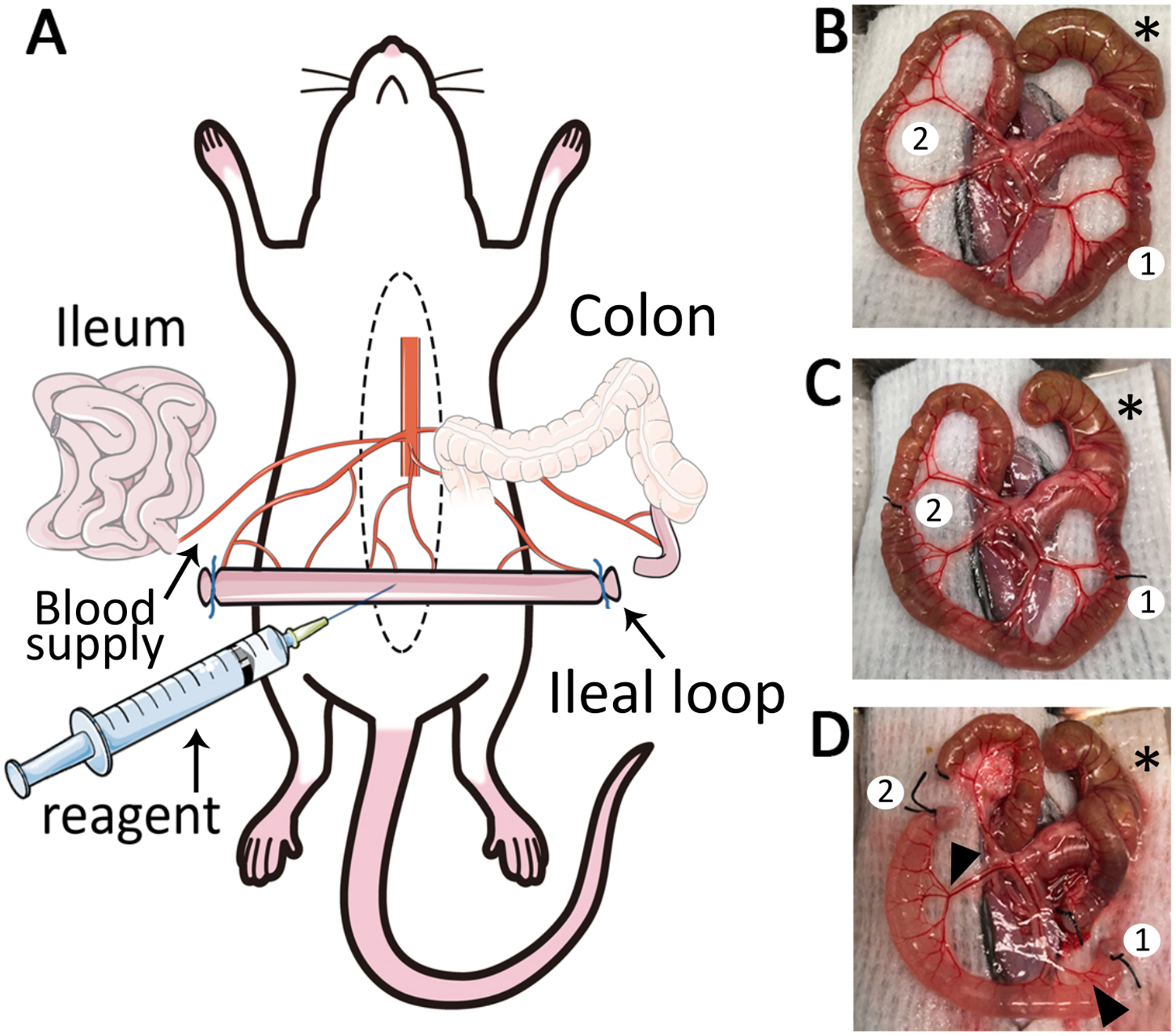
Figure 1: The ileal loop model.

(A) Schematic overview of the ileal loop model. Median laparotomy is performed on mice under anesthesia and placed on a temperature-controlled surgery board. (B) Exteriorization of the caecum (*), ileum and mesentery. Two adequate sites for ligation are identified (1,2). (C) Isolate a segment of 4 cm length: the first ligature (1) is placed close to the ileo-caecal junction and a second ligature (2) is placed 4 cm away from the first ligature. (D) Two small incisions are made in the mesentery (1, 2) to create a 4 cm length ileal loop. After removal of luminal content and ligation of cut-ends, reagents such as fluorescent markers and chemoattractants can be injected into the lumen. The ileal loop is well vascularized (black arrowheads).
