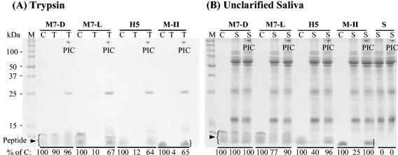
FIG. 2.
Stability of MUC7 12-mer-d (M7-D), MUC7 12-mer-l (M7-L), Hsn5 12-mer (H5), and magainin-II (M-II) in (A) trypsin and (B) unclarified saliva. Peptides were incubated with trypsin containing PIC or no PIC for 0.5 h or saliva (10 μl) for 1 h. The samples were analyzed by SDS-13% PAGE. After staining, the gel was analyzed for peptide degradation with a GS-700 imaging densitometer. The degradation was estimated by the equation [1 − (density of test band/density of control band)] × 100%. C, each peptide alone as a control; T, trypsin plus peptide; T+PIC, trypsin containing PIC plus peptide; S, saliva plus peptide; S+PIC, saliva containing PIC plus peptide. The most intense bands (the highest molecular mass) in the control (C) lanes represent full-length peptides, and the other bands (less intense) represent truncated peptides; for more detail, see the text. Densitometry data of control peptides and peptides remaining after incubation with trypsin or saliva are indicated at the bottom of each lane in each panel.

