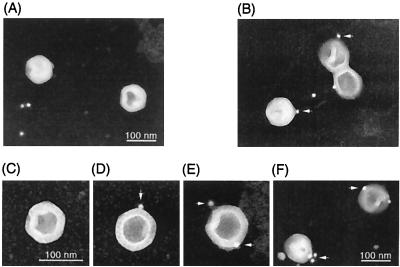
FIG. 2.
Electron microscopic immunostaining. B capsids were purified by gradient ultracentrifugation. B capsids were adsorbed to carbon grids and fixed in 2% glutaraldehyde. The grids were incubated with UL25 antiserum (B, D, E, and F) or normal mouse serum (A and C) and washed. Following incubation with 10-nm-gold-conjugated protein A, capsids were stained with uranyl acetate and examined by transmission electron microscopy. Arrows indicate gold particles on capsid vertices. Images of negative film are shown.

