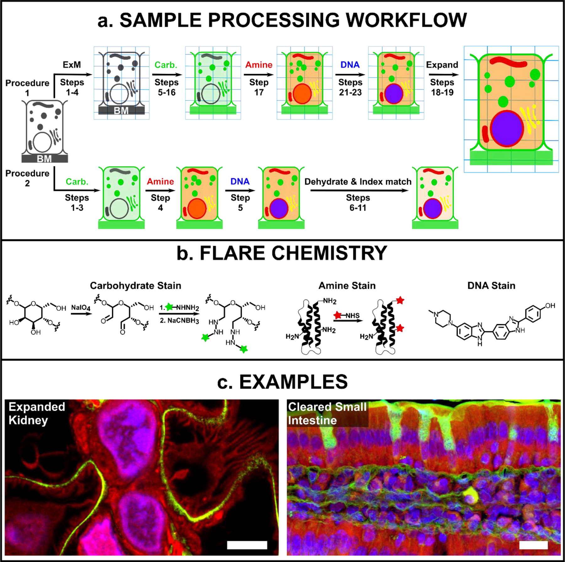
Fig. 1. Overview of FLARE for fluorescent labelling of biological samples.

(a) General schematic for FLARE staining and sample processing workflow for ExM (procedure 1) and cleared-tissue microscopy (procedure 2), where BM is the basement membrane. (b) Details of FLARE chemistry. Carbohydrates are oxidized to aldehydes using sodium periodate, coupled to hydrazide-functionalized dyes, and stabilized by reduction with sodium cyanoborohydride. N-hydroxysuccinimide (NHS)-functionalized dyes are used to label amine groups on proteins. DNA is labeled non-covalently using standard DNA-labeling dyes such as Hoechst. (c) Example confocal microscopy images of expanded mouse kidney (100 μm thickness) and optically-cleared mouse small intestine tissues (100 μm thickness) that have been stained by FLARE, where carbohydrates are green, accessible amines (on proteins) are red, and DNA is blue. Scale bars are 3 μm (left, pre-expansion units) and 10 μm (right).
