
Fig. 10. Demonstration of expansion uniformity using a MAP-based protocol.
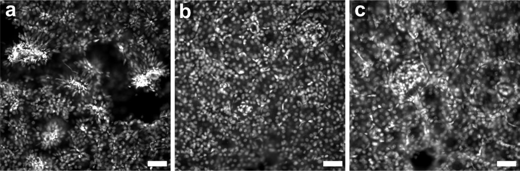
Epifluorescence images showing nuclei (DNA) stained by Sybr Green in expanded 100 μm thick mouse kidney tissue sections that were homogenized using a MAP-based protocol at three time points. (a) Incomplete homogenization leads to abundant tears (empty areas) and under-expanded regions (high-density clusters) exhibiting relatively small or abnormally stretched nuclei. (b) Extending the homogenization period (~5 times longer than in panel a) leads to uniform sample expansion that lacks obvious distortions in the expanded state. (c) A further extension of the homogenization period (~25 times longer than in panel a) does not cause a larger expansion factor or damage DNA. Scale bars 200 μm (a,b,c) in pre-expansion units.