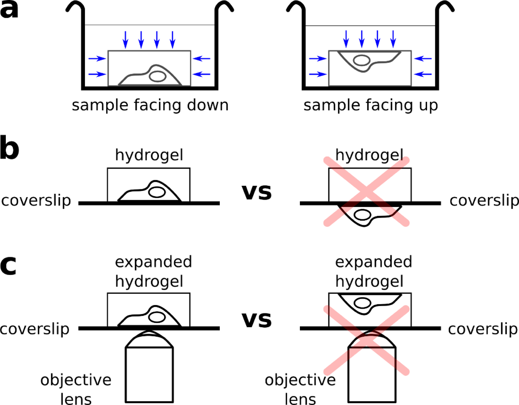
Fig. 11. Proper sample orientation during staining and image acquisition.

(a) If the cell sample is facing down (left), the dyes often have lower accessibility to the sample (blue arrows), resulting in dimmer stains. If the cell sample is facing up (right), the dyes have higher accessibility to the sample (blue arrows), resulting in brighter stains. (b) Hydrogel polymerization on the side coated with cells (left) vs polymerization on the incorrect side (right). If polymerization is performed on the side without the cells, then the subsequent hydrogel would not have any cells embedded, resulting in no signal under the microscope. (c) Expanded hydrogel mounted on poly-lysine coated coverslip, where cell sample is within the working distance of the objective (left) and the cell sample on the opposite side of coverslip (right). If the hydrogel is mounted as seen on the right, the cell sample might not be within the working distance of the objective lens.
