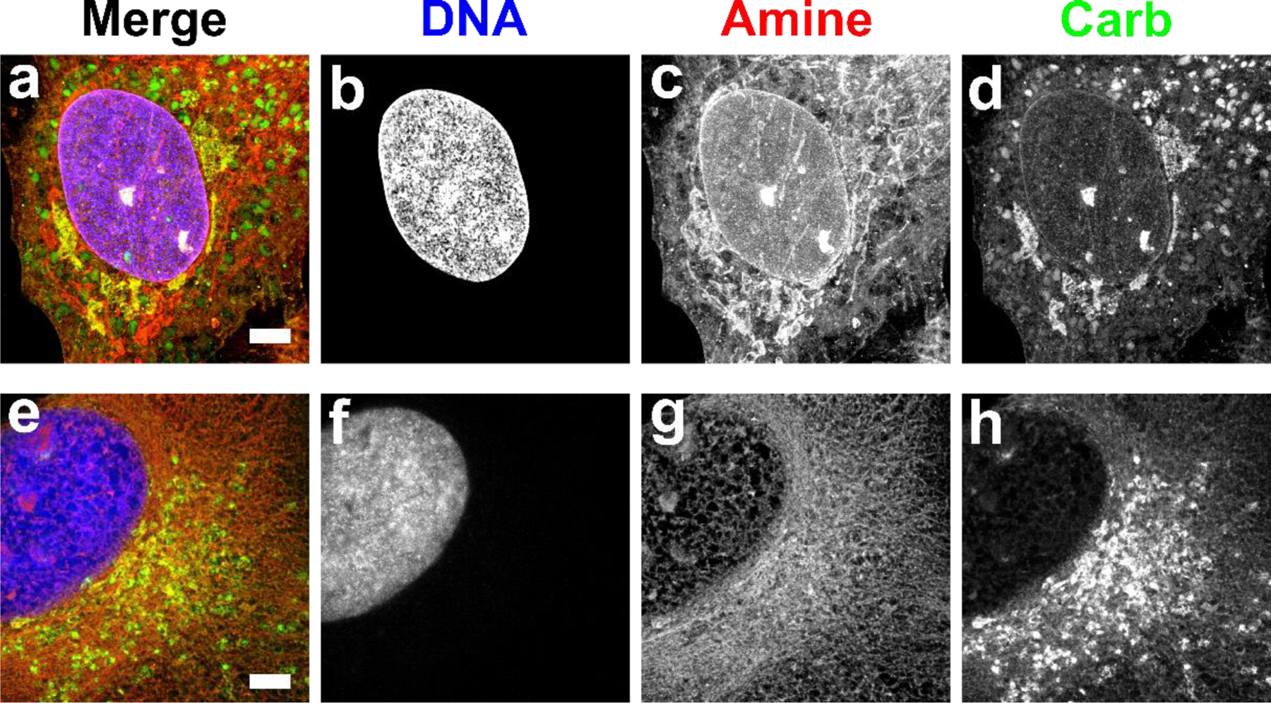
Fig. 3. Comparison of fixation conditions for expanded RPE cells stained by FLARE.

Red corresponds to amine stain, green corresponds to carbohydrate stain, and blue corresponds to nuclear stain. (a-d) Fixation with paraformaldehyde and glutaraldehyde generally preserved membrane-bound organelles and other structures while (e-h) fixation with glyoxal showed substantial perturbations. Scale bar (pre-expansion): 3 µm (a-h). Panels a-d were adapted from Mao et al.3 Science Advances. AAAS Publishing Group.
