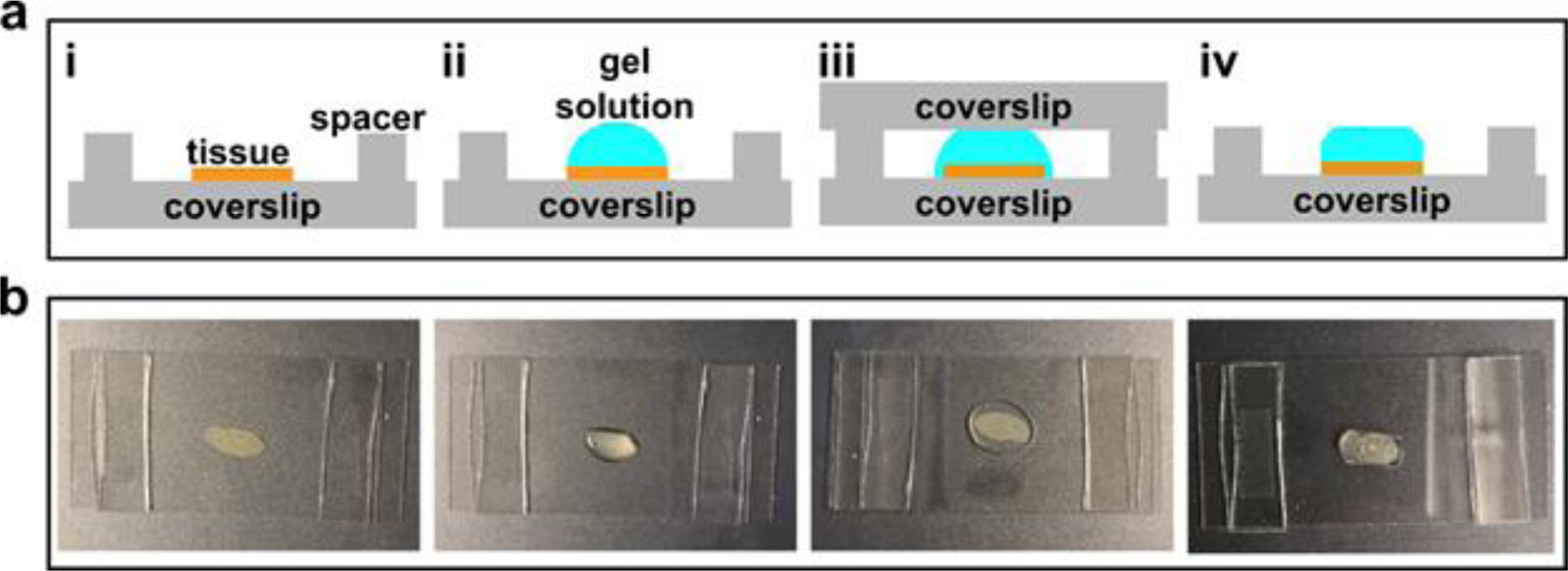
Fig. 6. Gelation chamber setup.

(a) Schematic of tissue sample gelation: i. spread a tissue sample on a coverslip (24mm × 50mm) and construct a spacer on each side using a stack of two #1.5 cover glass (~160 μm each); ii. Add a few drops of the freshly prepared gel solution to cover the entire sample area; iii. Place another coverslip (24 mm × 50 mm) on the top; iv. Once the gel has formed, peel off the top glass slide and trim off the excess gel. (b) Step-by-step photos correspond to i-iv in panel (a).
