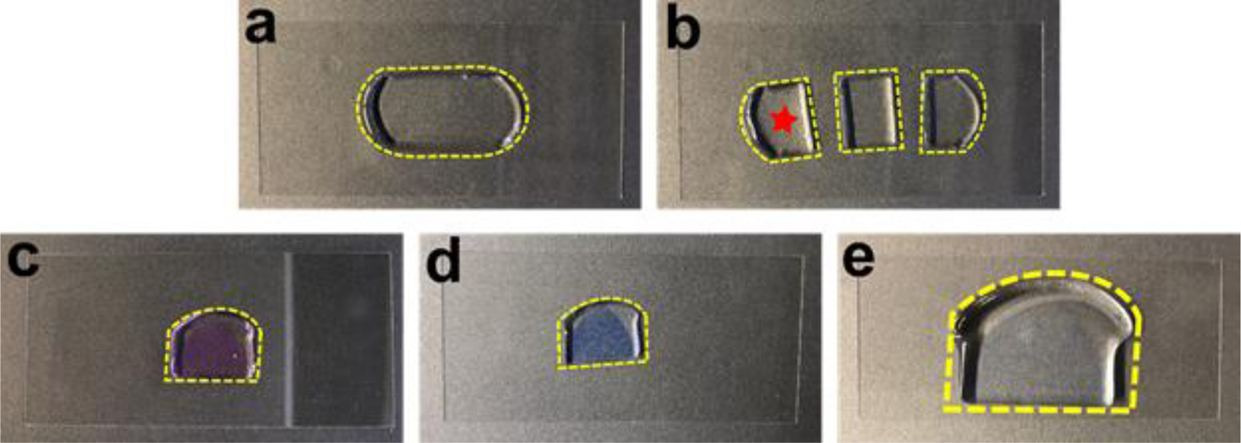
Fig. 7. A step-by-step illustration of the FLARE procedure for the expanded sample.

(a) The gel embedded tissue section shown in Figure 6 partially expanded after the denaturation and became transparent. (b) The tissue section was cut into three pieces. One piece (star) was used for FLARE staining and the other two were saved for other purposes. (c) The tissue section was stained pink after using ATTO 565 hydrazide for the carbohydrate stain. (d) Subsequently, the amine stain performed by using ATTO 647N-NHS ester made the tissue section light violet. (e) The tissue section was slightly colored even after the full expansion, demonstrating the characteristic of intense labeling of FLARE. The gel was laying down on a coverslip (24 mm × 50 mm) while taking step-by-step photos.
