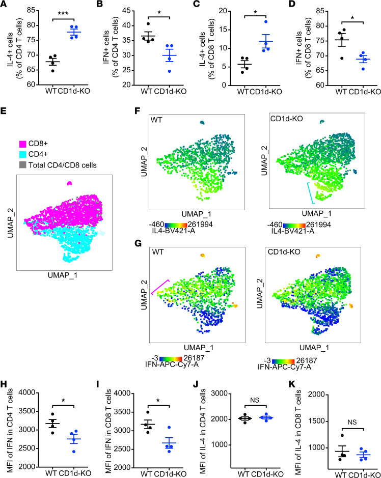
Figure 6. CD1ddep NKT cells promote Th1-associated cellular immunity.
The frequency of IL-4– or IFN-γ–producing CD4+ and CD8+ T cells in the LNs of WT and CD1d-KO mice was determined by intracellular staining, 3 days after s.c. DENV2 infection (n = 4). Percentages of (A) IL-4–producing CD4+ T cells (NK1.1–CD3+CD4+IL-4+), (B) IFN-γ–producing CD4+ T cells (NK1.1–CD3+CD4+IFN-γ+), (C) IL-4–producing CD8+ T cells (NK1.1–CD3+CD8+IL-4+), and (D) IFN-γ–producing CD8+ T cells (NK1.1–CD3+CD8+ IFN-γ+) are shown. (E) UMAP analysis showing CD4 and CD8 populations in infected dLNs. (F) A heatmap presentation of increased density of IL-4–expressing cells in CD1d-KO mice compared with WT in the CD4 compartment. Blue bracket indicates region of interest with increased density of IL-4hi cells. (G) Conversely, increased intensity for IFN-γ expression in WT mice compared with CD1d-KO mice in the CD8 compartment. Pink bracket indicates region of interest with IFN-γhi cells. (H and I) MFI of IFN-γ in CD4+ and CD8+ T cells indicates increased expression of IFN-γ in both CD4 and CD8 compartments in WT but not CD1d-KO mice. (J and K) MFI of IL-4 did not differ in cell types. n = 4 mice for each group; error bars represent mean ± SEM. *P < 0.05; ***P < 0.001, Student’s unpaired t test. During DENV infection, CD1ddep NKT cells are required for the optimal production of the Th1 cytokine IFN-γ by CD4+ and CD8+ T cells, and their absence causes a Th1/Th2 imbalance.

