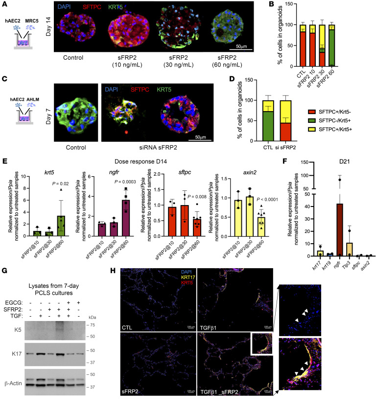
Figure 5. sFRP2 promotes BC differentiation of human AEC2s.
(A) Immunofluorescence for SFTPC and KRT5 of AEC2-derived organoids cocultured with MRC5 cells treated with sFRP2 for 14 days. Representative of n = 5 biological replicates. The experiment was performed in 3 technical triplicates, and data from 3 technical replicates are counted as 1 biological replicate. Original magnification, ×200. (B) Percentages of SFTPC+KRT5–, SFTPC+KRT5+, and SFTPC–KRT5+ cells in day-14 AEC2s plus MRC5 organoids treated with sFRP2. (C) Immunofluorescence of AEC2-derived organoids cocultured with AHLM after sFRP2 silencing. Original magnification, ×200. (D) Percentages of SFTPC+KRT5–, SFTPC+KRT5+, and SFTPC–KRT5+ cells in day-7 AEC2s plus AHLMsfrp2neg organoids. Data are presented as means of n = 4 biological replicates. (E) Levels of expression of krt5, ngfr, sftpc, and axin2 mRNA in EPCAM+ cells isolated from day-14 AEC2s plus MRC5 organoids treated with sFRP2. n = 3 biological replicates for sFFRP2 (10 and 30 ng/ml) and n = 4–7 biological replicates for sFRP2 (60 ng/ml). (F) Expression of genes characteristic for BCs in EPCAM+ cells isolated from day-21 AEC2s plus MRC5 organoids treated with sFRP2 (60 ng/ml). n = 2 biological replicates. (G and H) PCLSs from nondiseased donors were cultured and treated with or without TGF-β1 (2 ng/ml), with or without sFRP2 (60 ng/ml), and with or without EGCG (1 μM) for 7 days. (G) Lysates were blotted for KRT5 and KRT17. n = 3 biological replicates. (H) Immunofluorescence of KRT5 and KRT17. Representative of n = 3 independent experiments. Original magnification, ×100. Region of interest is presented as an insert (white rectangle) to show elongation of nuclei (DAPI) and cell morphology, outlined as a dotted line in insert as indicated by white arrowheads. Statistical significance was determined by mixed-effects analysis followed by Tukey’s multiple-comparisons test (B and D), Dunnett’s multiple comparisons test (E), and 2-tailed t test (F). P values are reported in Supplemental Table 4 for B and D.

