. 2024 Aug 17;16(8):e67050. doi: 10.7759/cureus.67050

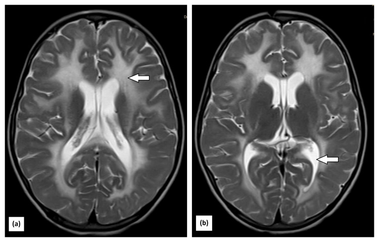
Figure 1. MRI showing (a) multiple ill-defined confluent T2 hyperintensities involving bilateral peri-ventricular white matter, deep white matter, and the bilateral fronto-parietal region. Tigroid appearance noted due to sparing of periventricular white matter; the bilateral external capsule and posterior limb of the internal capsule are involved; and subtle diffusion restriction in the splenium of the corpus callosum and along the lateral margins of white matter hyperintensity in both cerebral hemispheres; (b) bilateral cerebellar hemispheres involving white matter. All these findings support the diagnosis of vanishing white matter disease.

Figure 1