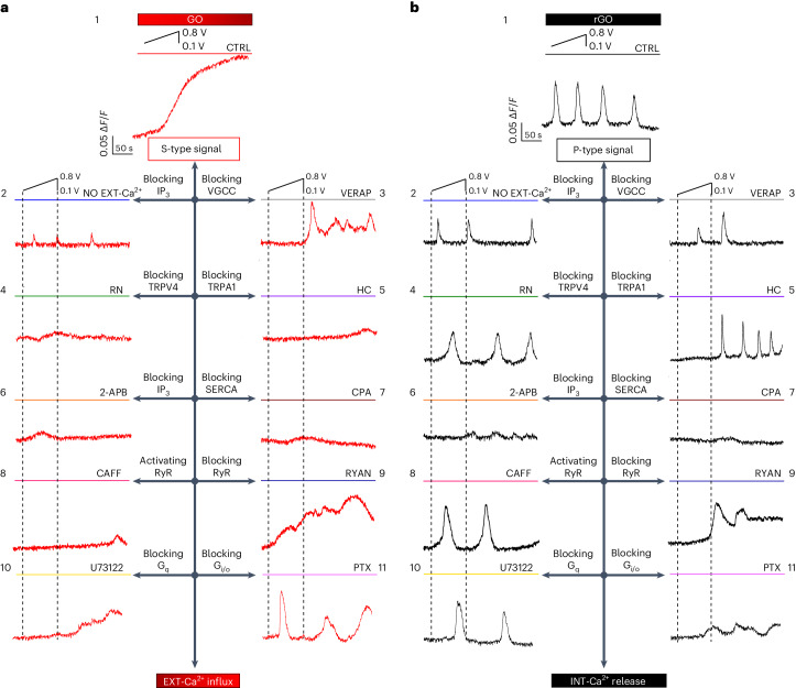
Fig. 2. Stimulation by GO/rGO coatings elicits distinct EXT-Ca2+ and INT-Ca2+ dynamics.
a,b, Representative traces of Ca2+ imaging observed after positive voltage bias stimulation of astrocytes, starting at time t ≈ 25 s from the beginning of the experiment (insets to panels 1) plated on GO–ITO- (a) and on rGO–ITO-coated electrodes (b). Different panels refer to the different conditions of the cells exposed to standard bath solution (CTRL, 1) and solution without extracellular Ca2+ (NO EXT-Ca2+, 2) and in the presence of VGCC inhibitor verapamil (VERAP, 25 μM, 3), TRPV4 inhibitor RN-1734 (RN, 10 μM, 4), TRPA1 inhibitor HC-030031 (HC, 40 μM, 5), IP3 receptor pathway inhibitor 2-aminoethoxy diphenyl borate (2-APB, 100 μM, 6), SERCA inhibitor cyclopiazonic acid (CPA, 10 μM, 7), RyR activator caffeine (CAFF, 20 mM, 8), RyR inhibitor ryanodine (RYAN, 50 μM, 9), Gq–PLC inhibitor U73122 (0.5 μM, 10) and Gi/o inhibitor pertussis toxin (PTX, 500 ng ml−1, 11).

