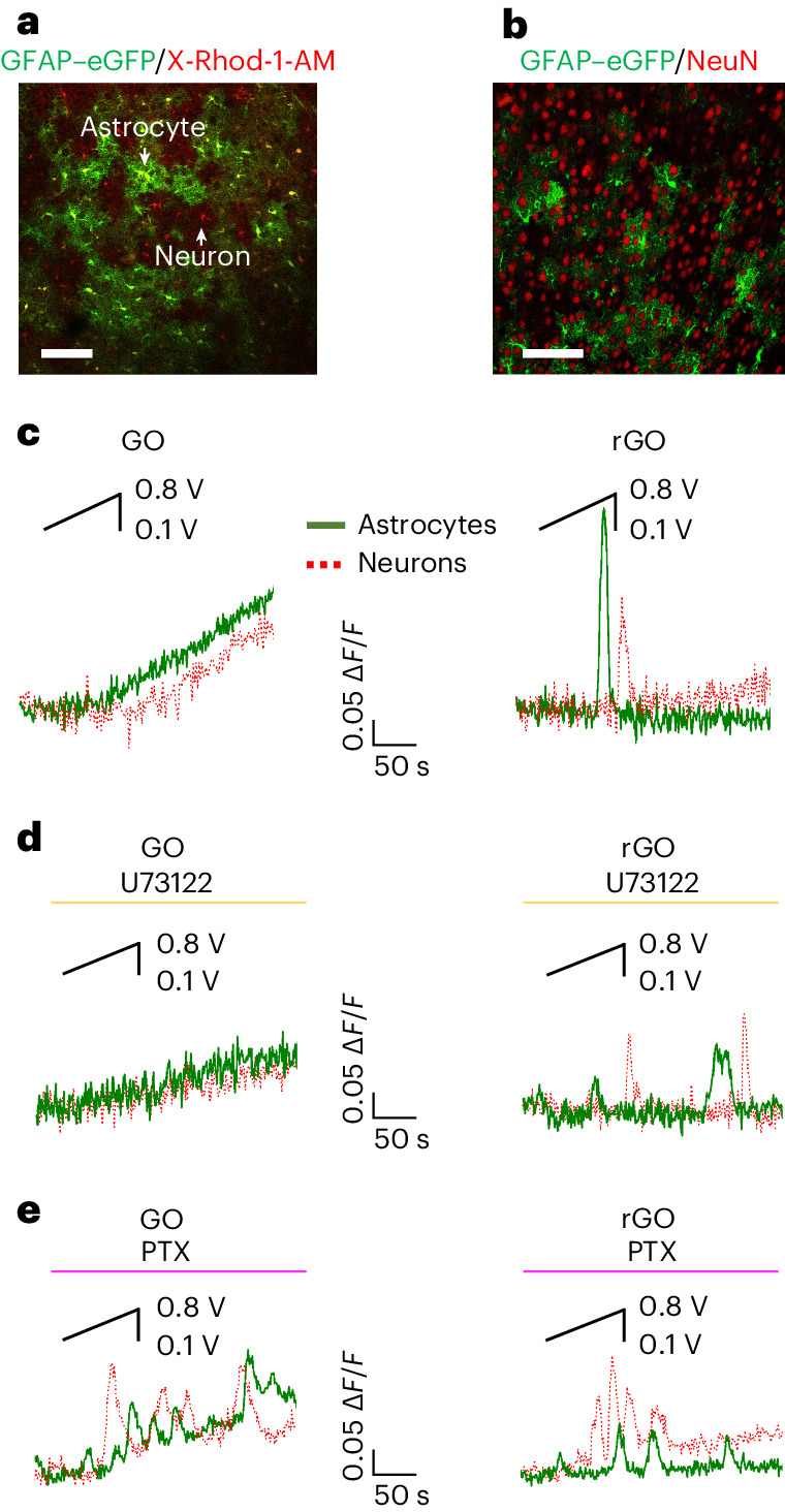
Fig. 5. Effects of GO and rGO stimulation on astrocyte and neuron GPCR signalling ex vivo.

a, Confocal fluorescence microscopy image of GFAP–eGFP/X-Rhod-1-AM-labelled cells revealing the co-presence of astrocytes (yellow cells) and neurons (red cells). b, Immunohistochemical image of GFAP–eGFP-labelled astrocytes (green cells) and neuronal cell protein marker (NeuN)-positive neurons (red cells) in brain slice. c–e, Representative traces of Ca2+ imaging experiments performed on brain slices lying on GO and rGO, analysed in neurons and astrocytes, recorded in control saline (c) and after exposure to U73122 (4 μM) (d) and Gi/o–GPCR inhibitor PTX (7.5 μg ml−1) (e).
