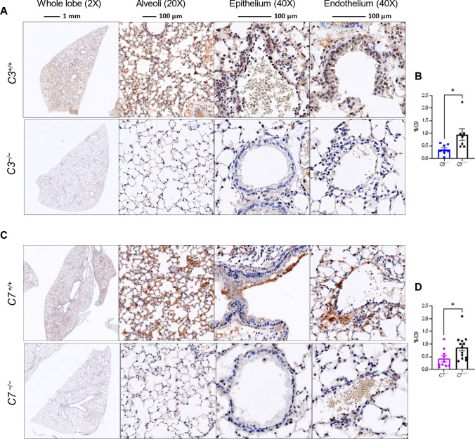
Fig. 7.
MAC deposition in the lungs as assessed by C9 staining. A–D Mouse lung sections were stained with an antibody against C9 (USBiological Life Sciences, 362359, Rabbit Anti-C9) used at a 1:1000 dilution. Representative images of the above groups showing whole lung lobe at magnification 2X, Alveoli at 20X, and epithelium and endothelium at 40X. A Representative image at 3DPI of lungs of C3 deficient and sufficient mice which were infected with MA30 at a dose of 5×10⁴ TCID50, and B quantification of C9 staining. C Representative image at 4DPI of lungs of C7 deficient and sufficient mice which were were infected with a dose of MA30 2.5×10⁴ TCID50, and D quantification of C9 staining. *p < 0.05 by two-tailed Student t-test

